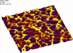
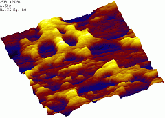
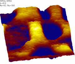
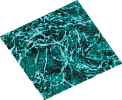
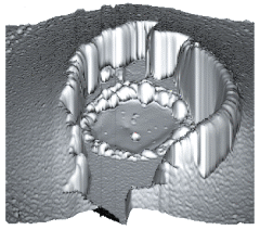
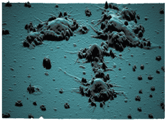
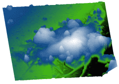
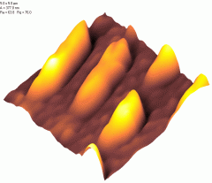
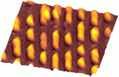
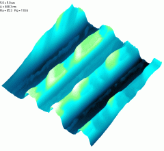
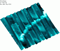
Contact mode, probes Mikromasch CSC12E
scan size 8.4x8.4 um
|
scan size 2.5x2.5 um |
scan size 1x1 um |
|
|
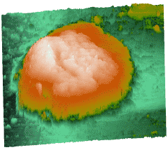
Tapping mode.
Probe Mikromasch NSC11B.
1.6x1.6 um. |
|
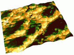
Mixed 3D image. 3D Frame: topography. Skin: phase shift. Tapping mode. Probe
Mikromasch NSC11B.
1.6x1.6 um. |
|
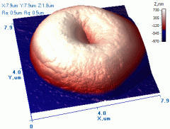
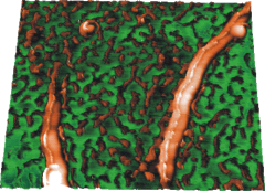
Tapping mode.
Probe Mikromasch NSC11B.
Scan size 7.9x7.9 um. |
|
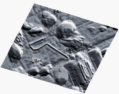
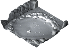
Contact mode.
Probe Mikromasch CSC38 B.
6.1x6.1 um. |
Contact mode.
Probe Mikromasch CSC38 B.
15.5x11.8 um. |
|
Contact mode.
Probe Mikromasch CSC38 B.
19.6x19.6 um. |
Contact mode.
Probe Mikromasch CSC38 B.
10.2x10.2 um. |
Contact mode.
Probe Mikromasch CSC38 B.
19.9x19.9 um. |
|
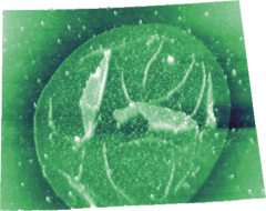
Contact mode. Probes Mikromasch CSC12E. 5x5 um |
Contact mode.
Probes Mikromasch CSC12E. 12x12 um |
|
Contact mode.
Probes Mikromasch CSC12F.
5x5 um |
Tappimg mode. Probes Mikromasch CSC12F. Scan size 5x5 um |
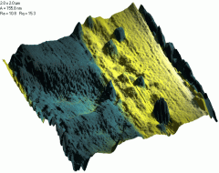
Joint image: Phase image as skin for 3D topography frame (blue - soft material,
yellow - harder substarte). Tapping mode. Probes Mikromasch CSC12F. 2x2 um |
|
Tapping mode.
Probe Mikromasch NSC11B.
Scan size 7.6x7.6 um. |
Tapping mode. Probe Mikromasch NSC11B.
Scan size [1.6x1.6 um] |
|
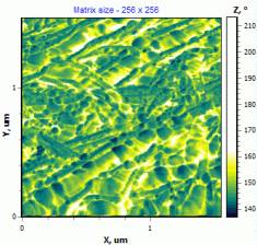
Topography image. Dynamic mode. Probes: Mikromasch NSC11A. Scan size [1.5x1.5
um] |
Phase shift image of the topography at left. Dynamic mode. Probes: Mikromasch
NSC11A. [1.5x1.5 um] |
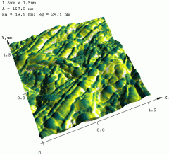
Joint image of previous two ones
(3D topography frame with skin according to phase image). |
|
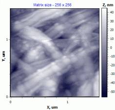
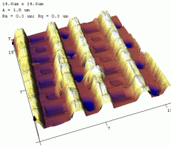
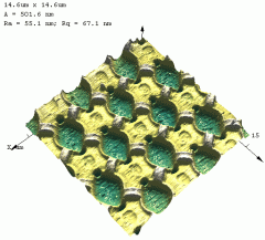
14.6x14.6 um. Contact mode |
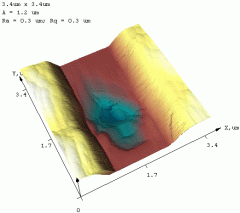
3.4x3.4 um. Contact mode |
|
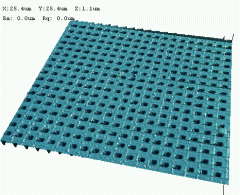
Topography image. Contact mode.
Probes: Mikromasch CSC38B. Scan size: 25.4x25.4 um |
|
Contact mode. 14.6x14.6 um |
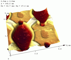
Contact mode. 4x4 um |
|
|
